Changes in the visual systems of newly diagnosed Parkinson’s disease patients may provide important biomarkers for the early detection and monitoring of the disease, according to a new study published online in the journal Radiology.
Changes in the visual systems of newly diagnosed Parkinson’s disease patients may provide important biomarkers for the early detection and monitoring of the disease, according to a new study published online in the journal Radiology.
“Just as the eye is a window into the body, the visual system is a window into brain disorders,” said lead researcher Alessandro Arrigo, M.D., a resident in ophthalmology at the University Vita-Salute San Raffaele of Milan, Italy.
Parkinson’s disease is a neurodegenerative condition caused by neuronal loss in several brain structures. Parkinson’s disease is characterized by tremors, rigidity or stiffness throughout the body, and impaired balance and coordination.
“Although Parkinson’s disease is primarily considered a motor disorder, several studies have shown non-motor symptoms are common across all stages of the disease,” Dr. Arrigo said. “However, these symptoms are often undiagnosed because patients are unaware of the link to the disease and, as a result, they may be under-treated.”
Read more at Radiological Society of North America
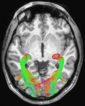
Image: Left and right optic radiation (OR) image in a representative subject overlaid onto a T1-weighted axial volume image of the same subject. OR images were obtained on the basis of diffusion-weighted volume images by means of constrained spherical deconvolution fitting and related tractography. Each bundle was automatically colored according to tract main directionality: red for left to right, green for anterior to posterior, and blue for inferior to superior. (Credit: Radiological Society of North America)


