Researchers at Uppsala University have developed a new method for very rapidly determining whether infection-causing bacteria are resistant or susceptible to antibiotics. The findings have now been published in the US journal Proceedings of the National Academy of Sciences (PNAS).
Researchers at Uppsala University have developed a new method for very rapidly determining whether infection-causing bacteria are resistant or susceptible to antibiotics. The findings have now been published in the US journal Proceedings of the National Academy of Sciences (PNAS).
Antibiotic resistance is a growing medical problem that threatens human health globally. One important contributory factor in the development of resistance is the incorrect use of antibiotics for treatment. Reliable methods to quickly and easily identify bacterial resistance patterns (Antibiotic Susceptibility Testing, AST) and provide the proper treatment from the start, i.e. right from the doctor’s appointment, are a solution to the problem. This has not been possible because existing antibiotic resistance tests take too long. Now, researchers at Uppsala have, for the first time, developed an antibiotic resistance test that is fast enough to enable a patient to take the right antibiotic home from the health centre straight after the first appointment. The test is primarily intended for urinary tract infections – a condition that, globally, affects approximately 100 million women a year and accounts for 25 per cent of antibiotic use in Sweden.
“We’ve developed a new method that allows determination of bacterial resistance patterns in urinary tract infections in 10 to 30 minutes. By comparison, the resistance determination currently in use requires one to two days. The rapid test is based on a new plastic microfluidic chip where the bacteria are trapped and methods for analysing bacterial growth at single-cell level,” says PhD student Özden Baltekin, who performed most of the experimental work.
Read more at Uppsala Universitet
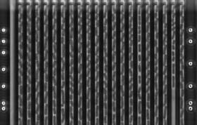
Image: This image shows Klebsiella pneumoniae growing in the microfluidic chip imaged in phase contrast. The bacteria are 0.003mm long and divide every 30 min. (Credit: Özden Baltekina, et al)


