A new study by Osaka University scientists shows that non-labeling multiphoton microscopy (NL-MPM) can be used for quantitative imaging of cancer that is safe and requires no resection, fixation or staining of tissues. The report is expected to simplify and reduce the time of cancer diagnosis and can be read in Scientific Reports.
A new study by Osaka University scientists shows that non-labeling multiphoton microscopy (NL-MPM) can be used for quantitative imaging of cancer that is safe and requires no resection, fixation or staining of tissues. The report is expected to simplify and reduce the time of cancer diagnosis and can be read in Scientific Reports.
To diagnose cancer and determine the proper treatment plan, pathologists depend on biopsies from the patient. The cutting, fixing and staining of the specimen generally provides reliable information, but these pathology-based diagnostic procedures require substantial time and cause tissue damage. Professor Masaru Ishii and his team of scientists have been exploring non-labeling microscopy methods as a means to observe and diagnose the cancer while preserving as much information as possible.
“MPM is an effective tool that enables the visualization of deep regions within living tissues and organs. We are developing NL-MPM to diagnose the malignancy of colorectal cancer,” he said.
Colorectal cancer has some ideal properties for the application of NL-MPM. Traditionally, microscopy of living organisms has depended on the attachment of a fluorescent dye to the target tissue. However, these dyes are often toxic, and it would be desirable to visualize human tissues without the need for additional labeling. Colorectal cancer particularly affects epithelial tissue, which emanates a sufficient signal in NL-MPM without the addition of any foreign dye, because chemicals natural to the tissue will emit autofluorescence.
Read more at Osaka University
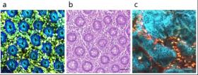
Image: These are images of "tissue diagnosis without cuttage". (a-b) Representative image of normal colorectal mucosal tissue by NL-MPM (a). It is easy to detect the characteristics of colorectal mucosa such as ductal structures in NL-MPM image, compared to conventional HE staining image (b). (c) Representative NL-MPM image of colorectal carcinoma tissue. It is easy to recognize the morphological features of colorectal carcinoma. (Credit: Osaka University)


