An estimated 5.5 million Americans live with Alzheimer’s disease, a type of dementia that causes problems with memory, thinking and behavior. Although treatments can slow the worsening of symptoms, scientists are still working to better understand the neurodegenerative disease so that curative and preventative medicines can be developed. A new imaging system could help speed new drug development by offering a better way to monitor the brain changes indicative of Alzheimer’s in mouse models of the disease.
An estimated 5.5 million Americans live with Alzheimer’s disease, a type of dementia that causes problems with memory, thinking and behavior. Although treatments can slow the worsening of symptoms, scientists are still working to better understand the neurodegenerative disease so that curative and preventative medicines can be developed. A new imaging system could help speed new drug development by offering a better way to monitor the brain changes indicative of Alzheimer’s in mouse models of the disease.
Mice genetically modified to exhibit characteristics of Alzheimer’s disease are a valuable tool for studying the disease’s biology and testing new drugs. Like people with the disease, the brains of these mice accumulate clumps of proteins known as senile plaques. Qingming Luo’s Visible Brain-wide Networks team at the Huazhong University of Science and Technology, China developed a system called cryo-micro-optical sectioning tomography (cryo-MOST) that improves the ability to image these senile plaques in the whole mouse brain.
“Studying the brain-wide distribution of senile plaques in mice will facilitate an understanding of how brain functions deteriorate during Alzheimer’s disease progression,” said Jing Yuan, a key member of the research team. “We hope that cryo-MOST will accelerate the development and evaluation of Alzheimer’s disease treatments.”
Read more at The Optical Society
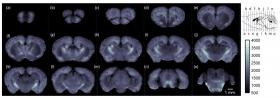
Image: Using cryo-MOST, the researchers acquired a brain-wide map of an Alzheimer mouse model showing that senile plaques had spread to the entire brain. The images are at 1-millimeter intervals from the olfactory bulb to the cerebellum. The specific locations are shown in the upper right insert. (Credit: Jing Yuan, Huazhong University of Science and Technology)


