
During operations, it can be difficult for surgeons to avoid severing crucial nerves because they look so much like other tissue. A new noninvasive approach that uses polarized light to make nerves stand out from other tissue could help surgeons avoid accidentally injuring nerves or assist them in identifying nerves in need of repair.
During operations, it can be difficult for surgeons to avoid severing crucial nerves because they look so much like other tissue. A new noninvasive approach that uses polarized light to make nerves stand out from other tissue could help surgeons avoid accidentally injuring nerves or assist them in identifying nerves in need of repair.
Although nerve injuries are a known complication for many types of surgery, surgeries involving the hand and wrist come with a higher risk because of the dense networks of nerves in this area. There are a few techniques available to help doctors identify nerves, but they have various limitations such as not providing real-time information, requiring physical contact with the nerve or requiring the addition of a fluorescent dye.
“We have shown that nerves can be distinguished in human tissue by detecting the interaction of light with the structure of nerves without the need for fluorescent markers or physical interaction,” said Kenneth Chin, a medical student at the Academic Medical Center (AMC), University of Amsterdam, Netherlands. “Using an intraoperative, noninvasive real-time method minimizes potential nerve damage, which can result in fewer negative consequences such as reduced function, loss of sensation or chronic pain.”
Cousins, Kenneth and Patrick Chin, developed the idea independently from any institute to use an optical technique known as collimated polarized light imaging (CPLi) to identify nerves during surgery. Kenneth later joined a research group led by Thomas van Gulik, a surgeon at the Academic Medical Center, and brought along a working prototype which has been further developed into a practical system that can be deployed in the operating room.
Read more at The Optical Society
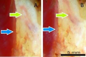
Image: The blue arrow highlights a vertical structure (A), which appears whiter after rotating the polarizers (B). The opposite occurs at the oblique structure (green arrow) indicating nerve tissue. (Credit: Kenneth Chin, Academic Medical Center)