Under a microscope, a cell’s cytoplasm can resemble a tiny underwater version of New York’s Times Square: Thousands of proteins swarm through a cytoplasm’s watery environment, coming together and breaking apart like a cytoskeletal flash mob.
Organelles such as mitochondria and lysosomes must traverse this crowded, ever-changing cytoplasmic space to deliver materials to various parts of a cell.
Now engineers at MIT have found that these organelles and other intracellular components may experience the surrounding cytoplasm as very different environments as they travel. For instance, a cell’s nucleus may “feel” the cytoplasm as a fluid, honey-like material, while mitochondria may experience it more like toothpaste.
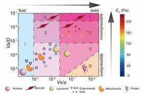
The team, led by Ming Guo, the Brit and Alex d'Arbeloff Career Development Assistant Professor in MIT’s Department of Mechanical Engineering, found that an organelle feels a certain resistance in cytoplasm, depending on that organelle’s size and the speed at which it moves through a cell. In particular, these characteristics determine how easily it can push against a cytoplasm’s surrounding water and move through its ever-changing web of cytoskeletal protein structures.
Read more at Massachusetts Institute of Technology
Image: A phase diagram of living mammalian cytoplasm, developed by MIT researchers, describes the type of material an organelle feels as it moves through cytoplasm, given its size and speed.
Image courtesy of the researchers


