A study of an unusual snapping turtle with one lung found shared characteristics with humans born with one lung who survive beyond infancy. Digital 3D anatomical models created by Emma Schachner, PhD, Assistant Professor of Cell Biology & Anatomy at LSU Health New Orleans School of Medicine, made the detailed research possible. The work is published in the December 2017 issue of The Journal of Anatomy, the cover of which features an image of the study’s 3D models.
A study of an unusual snapping turtle with one lung found shared characteristics with humans born with one lung who survive beyond infancy. Digital 3D anatomical models created by Emma Schachner, PhD, Assistant Professor of Cell Biology & Anatomy at LSU Health New Orleans School of Medicine, made the detailed research possible. The work is published in the December 2017 issue of The Journal of Anatomy, the cover of which features an image of the study’s 3D models.
“These shared traits include an enlarged single lung with a more homogenous distribution of respiratory parenchyma (the gas exchanging tissues), an opposing bronchus that ends where the opposite lung should be and malformations of the spine (such as scoliosis),” notes Dr. Schachner. “It is possible that similar genetic mutations are at play in both this turtle and in humans with this condition.”
The turtle was found in Minnesota and brought to the Wildlife Rehabilitation Center of Minnesota because of a bizarre shell deformity. When the single lung was discovered, Senior Veterinarian Renee Schott, DVM, contacted Schachner and co-author Dr. Tyler Lyson through their previous work on turtle lungs. Although the common snapping turtle has been well-studied, very little is known about developmental abnormalities and soft tissue pathologies of turtles and other reptiles.
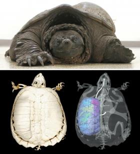
Using computed tomography (CT) and microCT imaging data and a pen tablet with special software, Schachner manually created 3D digital models of the areas of interest in both the live turtle and normal turtle specimens for comparison. She created solid 3D representations of the negative spaces within the lungs – the bronchial tree, the lung surface and the skeleton.
Read more at Louisiana State University Health Sciences Center
Image: This is a live turtle and 3-D model. (Credit: LSU Health New Orleans)


