
Researchers have developed a new device to map the brain during surgery and distinguish between healthy and diseased tissues. The device provides higher resolution neural readings than existing tools used in the clinic and could enable doctors to perform safer, more precise brain surgeries.
The device is an improved version of a clinical tool called an electrode grid, which is a plastic or silicone-based grid of electrodes that is placed directly on the surface of the brain during surgery to monitor the activity of large groups of neurons. Neurosurgeons use electrode grids to identify which areas of the brain are diseased in order to avoid damaging or removing healthy, functional tissue during operations. Despite their wide use, electrode grids have remained bulky and have not experienced any major advances over the last 20 years.
The new electrode grid, developed by a team of researchers at the University of California San Diego and Massachusetts General Hospital, is about a thousand times thinner — 6 micrometers versus several millimeters thick — than clinical electrode grids. This allows it to conform better to the intricately curved surface of the brain and obtain better readings. The new electrode grid also contains a much higher density of electrodes — spaced 25 times closer than those in clinical electrode grids — enabling it to generate higher resolution recordings.
“Our goal is to develop a tool that can obtain more reliable information from the surface of the brain,” said electrical engineering professor Shadi Dayeh, who co-led the study with neuroscience professor Eric Halgren and electrical engineering professor Vikash Gilja, all at UC San Diego. The project was funded by the Center for Brain Activity Mapping (CBAM) at UC San Diego and brought together experts from multiple fields, including neurosurgeons, neuroscientists, electrical engineers, materials scientists and experts in systems integration. Researchers published their work on May 12 in Advanced Functional Materials.
Read more at University of California - San Diego
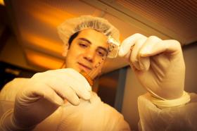
Photo: The PEDOT:PSS electrode grid is a new brain mapping device that can be used during brain surgery. (Credit: David Baillot/UC San Diego Jacobs School of Engineering)