
Optical Mammography, or OM, which uses harmless red or infrared light, has been developed for use in conjunction with X-rays for diagnosis or monitoring in cases demanding repeated imaging where high amounts of ionizing radiation should be avoided. At the OSA Biophotonics Congress: Biomedical Optics meeting, held 3-6 April in Hollywood, Florida, USA, researchers from Milan, Italy, will report an advance in instrument development that increases the sensitivity of OM by as much as 1000-fold.
Optical Mammography, or OM, which uses harmless red or infrared light, has been developed for use in conjunction with X-rays for diagnosis or monitoring in cases demanding repeated imaging where high amounts of ionizing radiation should be avoided. At the OSA Biophotonics Congress: Biomedical Optics meeting, held 3-6 April in Hollywood, Florida, USA, researchers from Milan, Italy, will report an advance in instrument development that increases the sensitivity of OM by as much as 1000-fold.
In 2017, an estimated 252,710 new cases of invasive breast cancer were diagnosed in women and 2,470 cases were diagnosed in men[i]. Many of these diagnoses are made using X-ray mammography. Although standard and widely used, X-ray imaging for breast cancer suffers from both low sensitivity (50-75%) and the use of ionizing radiation that cannot be considered completely safe.
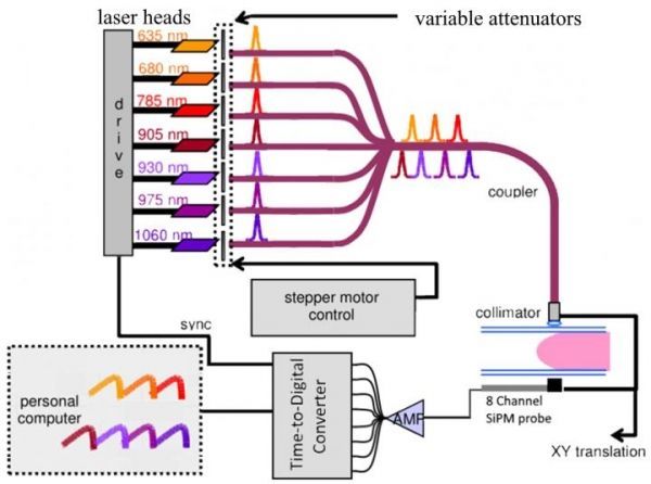
The newly-developed instrument replaces two photomultiplier tubes (PMTs) of existing instruments with an eight-channel probe involving silicon photomultipliers (SiPMs) and a multichannel time-to-digital converter. These changes eliminate a time-wasting pre-scan step that was required to avoid damage to the PMTs. In addition to increased sensitivity, the new instrument is both more robust and cheaper.
While X-ray mammography is widely used and is still the recommended method for routine screenings, its use is limited by the patient’s age, weight or body mass index, the breast tissue itself, whether or not hormone replacement therapy is being used, and other issues. In addition, its accuracy -- particularly when used in younger women -- has been called into question. Other imaging techniques, such as MRI and ultrasound, are sometimes suggested, but neither is an effective replacement for X-ray mammography.
Read more at The Optical Society
Image: Schematic Diagram for OM Instrument: Seven pulsed lasers sequentially illuminate the compressed breast; transmitted light is detected by the 8-channel SiPM probe and the TDC acquires the signal. (Credit: Edoardo Ferocino)