
Angiogenesis, the formation of new blood vessels, is essential for tumor growth. A new study reported in The American Journal of Pathology describes a vascular stabilization biomarker that can visualize blood vessel activity, thus optimizing the timing of anticancer therapies including anti-angiogenics.
Angiogenesis, the formation of new blood vessels, is essential for tumor growth. A new study reported in The American Journal of Pathology describes a vascular stabilization biomarker that can visualize blood vessel activity, thus optimizing the timing of anticancer therapies including anti-angiogenics.
Combination therapy using angiogenesis inhibitors and anticancer drugs can improve drug delivery into tumor tissues and prolong progression-free survival. “Vascular normalization by angiogenesis inhibitors, such as vascular endothelial growth factor (VEGF) signaling inhibitors, is a promising method for improvement of chemotherapy. However, it is unclear how we can recognize the ‘window of opportunity’ for the tumor vascular normalizing period for effective timing of anticancer drug treatment. Therefore, biomarkers delineating this window are essential,” explained Nobuyuki Takakura, MD, PhD, Professor, Department of Signal Transduction, Research Institute for Microbial Diseases, Osaka University, Osaka, Japan.
Researchers showed that active proliferating vascular endothelial cells (ECs) in mice could be distinguished from dormant ones. They measured the promoter activity of DNA replication factor partner of Sld5-1 (PSF1; official name GINS1) in ECs using enhanced green fluorescent protein (EGFP) that allows visualization of gene activity as fluorescence.
No EGFP signals were observed in normal adult skin vasculature, which was expected as normal skin ECs are dormant. However, after subcutaneous injection of tumor cells, some ECs in and near the tumor shifted to EGFP-positivity. PSF1 promotor activity was also found to correlate well with tumor cell growth. ECs that were high in EGFP expression were larger and had greater intracellular complexity than cells that were EGFP negative. “Our data showed that PSF1-promotor-EGFP mice may be utilized to visualize proliferating ECs by their EGFP expression,” commented Dr. Takakura.
Read more at Elsevier
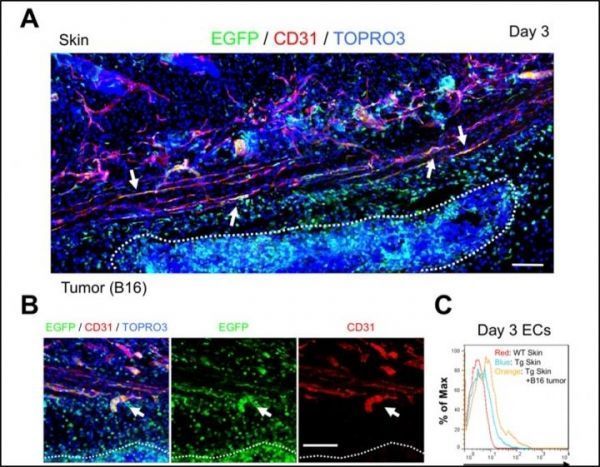
Image: Initiation of tumor angiogenesis up-regulates PSF1 promoter activity in endothelial cells (ECs). A: Skin vasculature 3 days after B16 melanoma cell injection into the mouse. White dashed line indicates localization of tumor. Arrows indicate EGFP-positive ECs around the tumor. Two pieces of images were put together and edited to one piece. B: Sprouting new capillary from preexisting blood vessels around the tumor. White dashed line indicates localization of tumor. Arrows indicate budding ECs that express EGFP and CD31 (red). C: Flow cytometric analysis of endothelial PSF1 promoter activity in skin tissues around tumors 3 days after tumor cell inoculation. PSF1 promoter activity in ECs is increased in the skin vasculature, with B16 tumor inoculation (orange) relative to skin from wild type (WT; red) or transgenic (Tg; blue) mice in the steady state. (Credit: The American Journal of Pathology)