
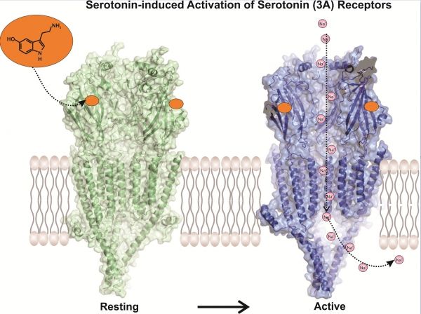
Serotonin (3A) receptors are common drug targets in the treatment of pain, gastrointestinal dysfunctions, and mood disorders yet little is known about their three-dimensional structure. Details about serotonin receptor structures could provide important clues to designing better drugs with less side effects. Now, a team of researchers from Case Western Reserve University School of Medicine have used high-powered microscopes to view serotonin activating its receptor for the first time. Images published in Nature reveal molecular details about the receptor that could improve drug design to treat a multitude of diseases.
Serotonin (3A) receptors are common drug targets in the treatment of pain, gastrointestinal dysfunctions, and mood disorders yet little is known about their three-dimensional structure. Details about serotonin receptor structures could provide important clues to designing better drugs with less side effects. Now, a team of researchers from Case Western Reserve University School of Medicine have used high-powered microscopes to view serotonin activating its receptor for the first time. Images published in Nature reveal molecular details about the receptor that could improve drug design to treat a multitude of diseases.
Serotonin receptors sit in cell membranes throughout the body, including the brain, stomach, and the associated nervous system. Drugs that inhibit serotonin receptors help control post-operative nausea, support cancer therapies, and are used to treat gastrointestinal conditions like irritable bowel syndrome. These inhibitors also find application as anti-depressants and promote attention and memory.
Read more at Case Western Reserve University
Image: Serotonin-induced activation of serotonin (3A) receptors. CREDIT: Case Western Reserve University