
Woods Hole Oceanographic Institution (WHOI) scientists are using advanced medical imaging techniques and diagnostic tools to reveal the internal structures of a wide range of marine animals.
Woods Hole Oceanographic Institution (WHOI) scientists are using advanced medical imaging techniques and diagnostic tools to reveal the internal structures of a wide range of marine animals. Most recently they worked on an international collaboration on mollusks: a diverse group of invertebrate sea creatures that includes snails, clams, mussels, squid, and octopuses.
The resulting images, made from digitally reconstructed radiographs (DRR), computed tomography (CT) scans, and magnetic resonance imaging (MRI), provide researchers with a non-destructive, non-invasive way to view the internal organs of animals in three dimensions—something that previously was only available by using conventional dissection methods.
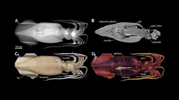
A paper published recently in the journal American Malacological Bulletin highlights stunning imagery from more than 20 scanned specimens ranging in size from a few millimeters to over one meter. The image above is of a Humboldt squid (Dosidicus), which is the largest of the mollusks imaged.
Read more at Woods Hole Oceanographic Institution
Image: Computed tomography (CT) images of an adult specimen of the cephalopod Dosidicus gigas. (A) Digitally reconstructed radiograph rendering, dorsal view with anterior facing right. (B) Virtual horizontal section at the level of digestive gland and gills. (C) Solid volume rendering showing external anatomical features. (D) Semi-transparent volume rendering exposing selected internal organs. (Images by Computerized Scanning and Imaging Facility, Woods Hole Oceanographic Institution)