
Many types of cancer could be more easily treated if they were detected at an earlier stage.
Many types of cancer could be more easily treated if they were detected at an earlier stage. MIT researchers have now developed an imaging system, named “DOLPHIN,” which could enable them to find tiny tumors, as small as a couple of hundred cells, deep within the body.
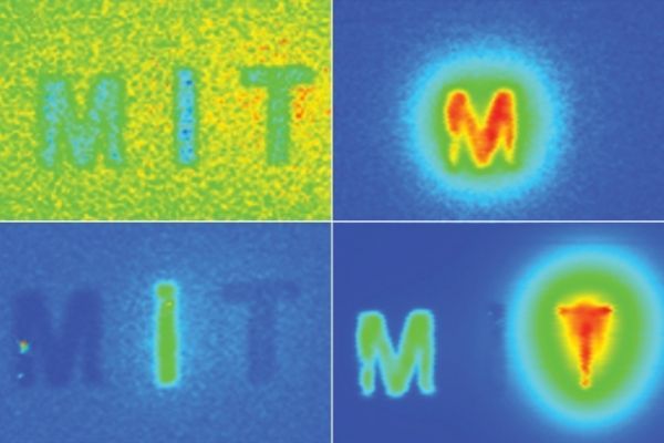
In a new study, the researchers used their imaging system, which relies on near-infrared light, to track a 0.1-millimeter fluorescent probe through the digestive tract of a living mouse. They also showed that they can detect a signal to a tissue depth of 8 centimeters, far deeper than any existing biomedical optical imaging technique.
The researchers hope to adapt their imaging technology for early diagnosis of ovarian and other cancers that are currently difficult to detect until late stages.
“We want to be able to find cancer much earlier,” says Angela Belcher, the James Mason Crafts Professor of Biological Engineering and Materials Science at MIT and a member of the Koch Institute for Integrative Cancer Research, and the newly-appointed head of MIT’s Department of Biological Engineering. “Our goal is to find tiny tumors, and do so in a noninvasive way.”
Read more at Massachusetts Institute of Technology
Image: MIT researchers have devised a way to simultaneously image in multiple wavelengths of near-infrared light, allowing them to determine the depth of particles emitting different wavelengths. (Image courtesy of the researchers)