
A simple MRI method that measures iron content can provide a more comprehensive picture of the consequences of stroke-related damage to the brain, according to a study published in Radiology.
A simple MRI method that measures iron content can provide a more comprehensive picture of the consequences of stroke-related damage to the brain, according to a study published in Radiology.
Strokes can result in significant impairments caused by infarction and long-term outcomes may be affected by delayed degeneration of brain areas away from the location of the stroke. One such area is the substantia nigra (SN).
“Overall, the SN is strongly involved in motor control, but also in regulation of emotions, cognition and motivation,” said study co-author Thomas Tourdias, MD, PhD, professor of radiology at the Centre Hospitalier Universitaire in Bordeaux, France. “Usually, stroke doesn’t directly affect the SN but, by interrupting circuits, stroke can induce secondary degeneration of that area.”
Prior to the new study, this secondary degeneration of SN had only been seen on post-mortem brain examinations. Imaging on a living person has not been able to capture the long-term degeneration, so for the new study, Dr. Tourdias and colleagues employed MRI R2* mapping to measure iron content in the brain.
Read more at Radiological Society of North America
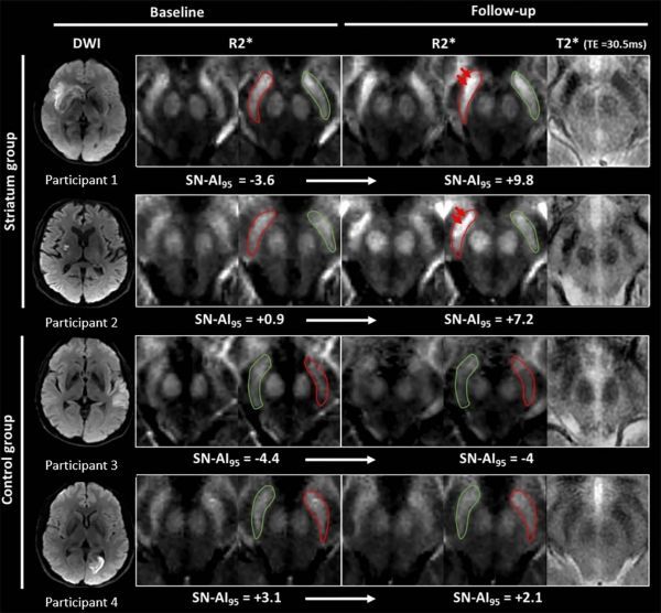
Image: Images show illustrative examples of visual R2? modifications within substantia nigra (SN) at baseline (24-72 h) and follow-up (1 y) in striatum (participants 1 and 2) and control groups (participants 3 and 4). (Credit: Radiological Society of North America)