
Using genetic tools in mice, researchers at Johns Hopkins Medicine say they have identified a pair of proteins that precisely control when sound-detecting cells, known as hair cells, are born in the mammalian inner ear. The proteins, described in a report published June 12 in eLife, may hold a key to future therapies to restore hearing in people with irreversible deafness.
“Scientists in our field have long been looking for the molecular signals that trigger the formation of the hair cells that sense and transmit sound,” says Angelika Doetzlhofer, Ph.D., associate professor of neuroscience at the Johns Hopkins University School of Medicine. “These hair cells are a major player in hearing loss, and knowing more about how they develop will help us figure out ways to replace hair cells that are damaged.”
In order for mammals to hear, sound vibrations travel through a hollow, snail shell-looking structure called the cochlea. Lining the inside of the cochlea are two types of sound-detecting cells, inner and outer hair cells, which convey sound information to the brain.
An estimated 90% of genetic hearing loss is caused by problems with hair cells or damage to the auditory nerves that connect the hair cells to the brain. Deafness due to exposure to loud noises or certain viral infections arises from damage to hair cells. Unlike their counterparts in other mammals and birds, human hair cells cannot regenerate. So, once hair cells are damaged, hearing loss is likely permanent.Read more at: Johns Hopkins Medicine
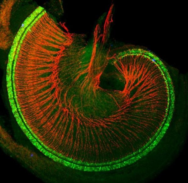
Mouse cochlea with hair cells shown in green and auditory nerves shown in red. (Photo Credit: Doetzlhofer lab)
Researchers Find Proteins That Might Restore Damaged Sound-Detecting Cells in the Ear
Typography
- Font Size
- Default
- Reading Mode