
For most people, getting an ultrasound is a relatively easy procedure: As a technician gently presses a probe against a patient’s skin, sound waves generated by the probe travel through the skin, bouncing off muscle, fat, and other soft tissues before reflecting back to the probe, which detects and translates the waves into an image of what lies beneath.
For most people, getting an ultrasound is a relatively easy procedure: As a technician gently presses a probe against a patient’s skin, sound waves generated by the probe travel through the skin, bouncing off muscle, fat, and other soft tissues before reflecting back to the probe, which detects and translates the waves into an image of what lies beneath.
Conventional ultrasound doesn’t expose patients to harmful radiation as X-ray and CT scanners do, and it’s generally noninvasive. But it does require contact with a patient’s body, and as such, may be limiting in situations where clinicians might want to image patients who don’t tolerate the probe well, such as babies, burn victims, or other patients with sensitive skin. Furthermore, ultrasound probe contact induces significant image variability, which is a major challenge in modern ultrasound imaging.
Now, MIT engineers have come up with an alternative to conventional ultrasound that doesn’t require contact with the body to see inside a patient. The new laser ultrasound technique leverages an eye- and skin-safe laser system to remotely image the inside of a person. When trained on a patient’s skin, one laser remotely generates sound waves that bounce through the body. A second laser remotely detects the reflected waves, which researchers then translate into an image similar to conventional ultrasound.
Read more at Massachusetts Institute of Technology
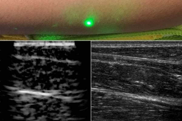
Image: A new ultrasound technique uses lasers to produce images beneath the skin, without making contact with the skin as conventional ultrasound probes do. The new laser ultrasound technique was used to produce an image (left) of a human forearm (above), which was also imaged using conventional ultrasound (right). Image courtesy of the researchers