
Using a combination of experimental and computational data, researchers discover paths to optimize pulses from highly intense X-ray beams.
Using a combination of experimental and computational data, researchers discover paths to optimize pulses from highly intense X-ray beams.
Scientists have long pursued the ability to see the structure of a single, free-form molecule at atomic resolution, what many call the “holy grail” of imaging. One potential method involves aiming extremely short, highly intense X-ray free-electron laser (XFEL) pulses at a sample material. But this ultrafast imaging technique also destroys its target, so time is of the essence.
Researchers at the U.S. Department of Energy’s (DOE) Argonne National Laboratory are advancing the effort with a combination of experiments and computer simulations, looking to understand how XFEL pulses interact with their targets. Recently, a team led by Argonne’s Atomic Molecular Optical Physics group in the Chemical Sciences and Engineering division pinpointed an important and often ignored parameter that can influence experiment outcomes: time. Their paper, “The role of transient resonances for ultra-fast imaging of single sucrose nanoclusters,” was recently published in the journal Nature Communications.
Read more at DOE / Argonne National Laboratory
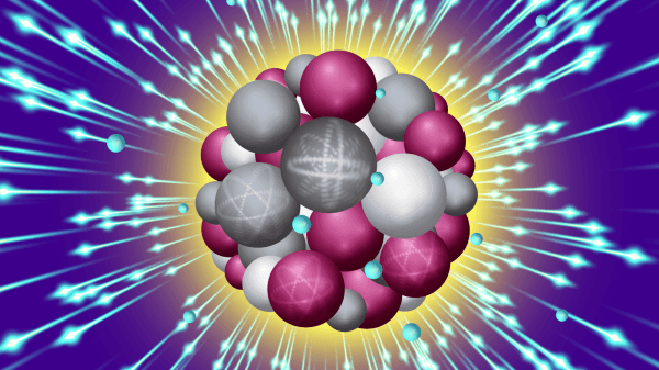
Image: An intense X-ray pulse scatters off a sucrose cluster (red, white, and gray spheres are oxygen, carbon and hydrogen atoms, respectively) resulting in ejected electrons (blue spheres) and structural deformation. (Image by Stacy Huang.)