
Patients with COVID-19 can have bowel abnormalities, including ischemia, according to a new study published in Radiology.
Patients with COVID-19 can have bowel abnormalities, including ischemia, according to a new study published in Radiology.
Several studies have evaluated the chest imaging findings in COVID-19, which helped improve understanding of how the disease affects the lungs. More recently, reports have documented that gastrointestinal symptoms, liver injury and vascular findings are common in these patients. However, abdominal imaging findings have not yet been widely reported. Imaging findings may help physicians understand abdominal manifestations in patients with the infection. Therefore, the authors of this study set out to explore abdominal imaging findings in patients with COVID-19.
Patients in the ICU More Likely to Develop Bowel Abnormalities
The retrospective study included 412 patients consecutively admitted to a single quaternary care center from March 27 to April 10, 2020, who tested positive for severe acute respiratory syndrome coronavirus 2 (SARS-CoV-2). The cohort included 241 men (58.5%) and 171 women (41.5%), with an average age of 57 years.
Read more at Radiological Society of North America
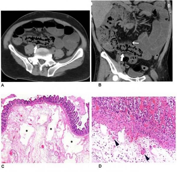
Image: Non-contrast axial (A) and coronal (B) CT performed in a 54-year-old man demonstrates pneumatosis cystoides intestinalis (arrows) in a long segment of ileum. Adjacent mesenteric congestion is also noted (thin arrow). Laparotomy demonstrated no frank bowel necrosis. Low power photomicrograph (H and E, 40x) of the ileum (C) shows ischemic degenerative changes of the mucosa with villous blunting (left) and withered crypts. There is marked submucosal edema with large empty spaces consistent with pneumatosis (*). High power view (H and E, 400x) (D) of the superficial submucosa shows arterioles with fibrin thrombi (arrowheads) beneath the damaged mucosa. (Credit: Radiological Society of North America)