
In recent weeks, a multisystem hyperinflammatory condition has emerged in children in association with prior exposure or infection to SARS-CoV-2.
In recent weeks, a multisystem hyperinflammatory condition has emerged in children in association with prior exposure or infection to SARS-CoV-2. A new case series published in Radiology examines the spectrum of imaging findings in children with the post-COVID-19 inflammatory condition known in the U.S. as Multisystem Inflammatory Syndrome in Children (MIS-C).
The array of findings includes airway inflammation and rapid development of pulmonary edema, coronary artery aneurysms, and extensive intra-abdominal inflammatory changes.
In April 2020, Evelina London Children’s Hospital in London, U.K., experienced a surge of children with a multi-system hyperinflammatory syndrome. The children had a variety of symptoms, including fever, headaches, abdominal pain, rash and conjunctivitis. Clinical features and lab findings shared some similarities to those of Kawasaki disease, Kawasaki-disease shock syndrome or toxic-shock syndrome, although atypical and more severe.
“Our hospital saw an unprecedented cluster of children presenting with MIS-C, a new hyperinflammatory syndrome in children related to the current COVID-19 pandemic — the recognition of which led to a national alert,” said the study’s lead author, Shema Hameed, MBBS, consultant pediatric radiologist at Evelina London Children’s Hospital.
Read more at Radiological Society of North America
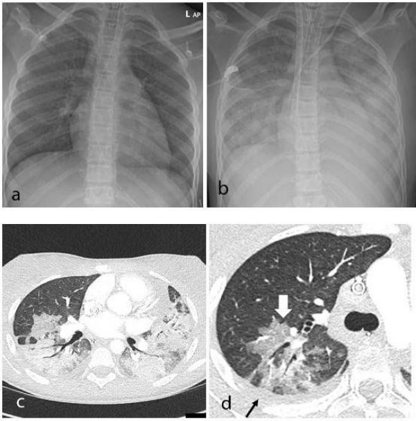
Image: Thoracic findings in a 15-year-old girl with Multisystem Inflammatory Syndrome in Children (MIS-C). (a) Chest radiograph on admission shows mild perihilar bronchial wall cuffing. (b) Chest radiograph on the third day of admission demonstrates extensive airspace opacification with a mid and lower zone predominance. (c, d) Contrast-enhanced axial CT chest of the thorax at day 3 shows areas of ground-glass opacification (GGO) and dense airspace consolidation with air bronchograms. (c) This conformed to a mosaic pattern with a bronchocentric distribution to the GGO (white arrow, d) involving both central and peripheral lung parenchyma with pleural effusions (black small arrow, d). (Credit: Radiological Society of North America)