
Most human diseases can be traced to malfunctioning parts of a cell — a tumor is able to grow because a gene wasn’t accurately translated into a particular protein or a metabolic disease arises because mitochondria aren’t firing properly, for example.
Most human diseases can be traced to malfunctioning parts of a cell — a tumor is able to grow because a gene wasn’t accurately translated into a particular protein or a metabolic disease arises because mitochondria aren’t firing properly, for example. But to understand what parts of a cell can go wrong in a disease, scientists first need to have a complete list of parts.
By combining microscopy, biochemistry techniques and artificial intelligence, researchers at University of California San Diego School of Medicine and collaborators have taken what they think may turn out to be a significant leap forward in the understanding of human cells.
The technique, known as Multi-Scale Integrated Cell (MuSIC), is described November 24, 2021, in Nature.
“If you imagine a cell, you probably picture the colorful diagram in your cell biology textbook, with mitochondria, endoplasmic reticulum and nucleus. But is that the whole story? Definitely not,” said Trey Ideker, PhD, professor at UC San Diego School of Medicine and Moores Cancer Center. “Scientists have long realized there’s more that we don’t know than we know, but now we finally have a way to look deeper.”
Read More: University of California - San Diego
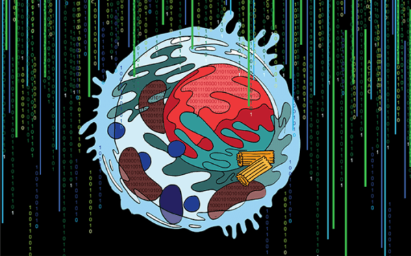
UC San Diego researchers introduce Multi-Scale Integrated Cell (MuSIC), a technique that combines microscopy, biochemistry and artificial intelligence, revealing previously unknown cell components that may provide new clues to human development and disease. (Artist’s conceptual rendering.) (Photo Credit: UC San Diego Health Sciences)