
Experts have long known that tumors arise from the migration of cancer cells across organs.
Experts have long known that tumors arise from the migration of cancer cells across organs. But observing the progression of those cancer cells in high resolution, and in turn being able to more effectively hinder their advances, has been a challenge for scientists. Now, a cutting-edge technique originally developed by Associate Professor Pavel Osten’s lab to image the mammalian brain has been adapted by postdoc Julian Taranda of Osten’s team and his CSHL colleagues in Professor Lloyd Trotman’s lab, in order to peek at prostate cancer cell migration in mice. The scientists have created a remarkably detailed 3D model of the tiny mouse prostate, allowing them to better observe cancer progression, with implications for effectively treating the disease in both mice and humans.
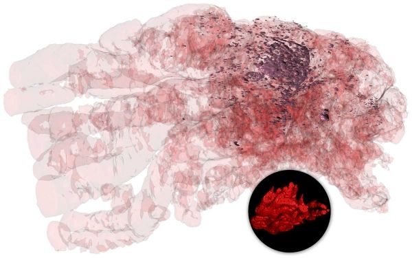
The imaging technique is called serial two-photon tomography (STPT). The two labs have used it to not only map in unprecedented detail the dense and complex structure that is a mouse prostate (inset image above, bottom right) but also to reveal the progression of the cancerous cells acting within (main image above).
Read more at Cold Spring Harbor Laboratory
Image Credit: Professor Pavel Osten’s lab