
Timing is everything when it comes to the embryonic development of pre-vertebrae divisions along the body of an embryo, according to researchers in Japan.
Timing is everything when it comes to the embryonic development of pre-vertebrae divisions along the body of an embryo, according to researchers in Japan. A new live-imaging technique in mouse cells suggests a specific ‘clock gene’ Hes7, oscillates at a time-delay to give rise to vertebrae, spinal column and occipital bone in vertebrates. The research, published in the journal Nature, sheds light on how cells’ internal communication is controlled and timed in normal development and which compounds are involved.
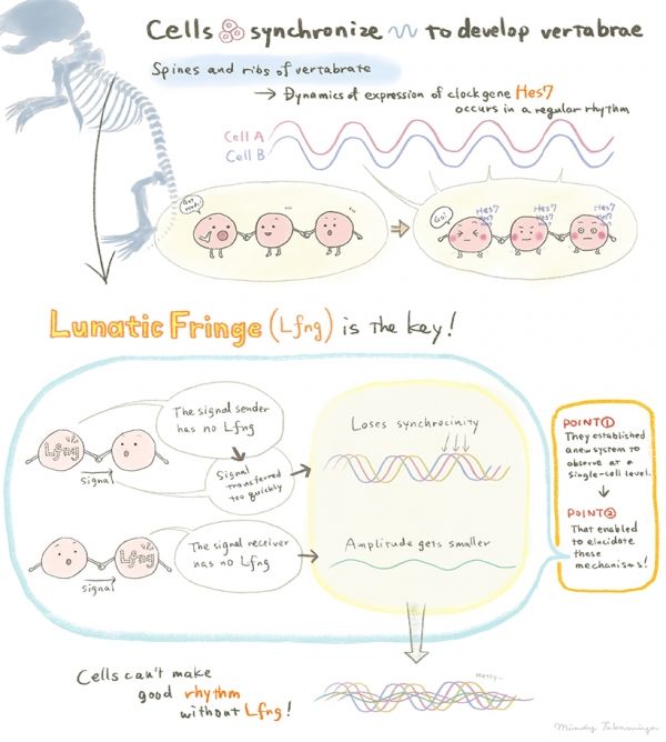
During embryonic development, the cells that will form nerve and connective tissue align themselves and begin to express relevant genes. The process occurs with a regular rhythm and is known to developmental biologists as the segmentation clock. The repetitive structures that are formed lie along the body axis and give rise to vertebrae and ribs.
Developmental biologist Ryoichiro Kageyama and colleagues at the Institute for Integrated Cell-Material Sciences (iCeMS) at Kyoto University wanted to understand the coordinated dynamics within cells. Until now this process has only been studied in snapshots – single images of cells but not using live images.
Read more at Kyoto University
Illustration by Izumi Mindy Takamiya. (CC BY 4.0)