
For one recent case, Dr. David Frumberg had to figure out how he was going to fix the left leg of his patient, a 14-year-old girl.
For one recent case, Dr. David Frumberg had to figure out how he was going to fix the left leg of his patient, a 14-year-old girl.
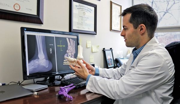
“She has an abnormal connection between these two bones, and she’s missing half of her tibia,” said Frumberg, assistant professor of orthopedics and rehabilitation. “Have you ever had pain on the outside of your foot? It’s awful, and she experiences that with every step.”
It’s a tricky case, and X-rays and CT scans weren’t giving him enough information to go on. To better understand what was going on, Frumberg went to the Center for Engineering Innovation & Design (CEID) to get a 3D-printed model of the joints. Having the model allowed him to hold the problem in his hand, turn it around, and help him see things he couldn’t before.
“For me, the model helps in so many ways,” he said. “It helps me come up with a plan, it helps me figure out the relationships between all the different complex deformities, and what needs to be repaired and what doesn’t.”
Read more at Yale University
Image Credit: Frumberg compares a 3D printed model to a traditional CT scan. CREDIT: Yale University