Spurred by the COVID-19 pandemic, Princeton researchers have developed a diagnostic tool to analyze chest X-rays for patterns in diseased lungs.
Spurred by the COVID-19 pandemic, Princeton researchers have developed a diagnostic tool to analyze chest X-rays for patterns in diseased lungs. The new tool could give doctors valuable information about a patient's condition, quickly and cheaply, at the point of care.
Jason Fleischer, professor of electrical engineering and the project's principal investigator, said he was inspired to create the tool after reading about COVID-19's devastating range of attacks. As hospitals have been overrun with patients, doctors have observed two basic types of lung damage, one more immediately life-threatening than the other. Treatment can differ between the types, so distinguishing the two could improve care and better allocate scarce resources.
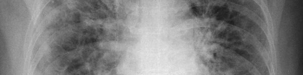
While current differentiation methods involve expensive and time-consuming procedures, such as computed tomography (CT) scans, Fleischer's machine learning model looks at a simple X-ray image and finds patterns that are too subtle even for the expert human eye. This tool would give doctors a new measure for determining the type and severity of COVID-19 pneumonia. And the process, on the ground, is simple.
Read more at Princeton University
Image: Princeton researchers have developed a diagnostic tool that uses AI to analyze chest X-rays for COVID-19 lung damage. The tool could help doctors triage patients and better allocate scarce resources. Image courtesy of the researchers. CREDIT: Princeton University