Measuring plant phenotypes, a term used to describe the observable characteristics of an organism, is a critical aspect of studying and improving economically important crops.
Measuring plant phenotypes, a term used to describe the observable characteristics of an organism, is a critical aspect of studying and improving economically important crops. Phenotypes central to the breeding process include traits like kernel number in corn, seed size in wheat, or fruit color in grape. These features are visible to the naked human eye but are in fact driven by microscopic molecular and cellular processes in the plant. Using three-dimensional (3D) imaging is a recent innovation in the plant biology sector to capture phenotypes on the “whole-plant” scale: from miniscule cells and organelles in the roots, up to the leaves and flowers. However, current 3D imaging processes are limited by time-consuming sample preparation and by imaging depth, usually reaching only a few layers of cells within a plant tissue. New research led by Christopher Topp, PhD, associate member at the Donald Danforth Plant Science Center, and Keith Duncan, a research scientist in his lab, have pioneered X-ray microscope technology to image plant cells, whole tissues, and even organs at unprecedented depths with cellular resolution. The work, supported by Valent BioSciences LLC and Sumitomo Chemical Corporation, was recently published in the scientific journal Plant Physiology, titled X-ray microscopy enables multiscale high-resolution 3D imaging of plant cells, tissues, and organs. This work will enable plant scientists globally to study above and below-ground traits at revolutionary clarity.
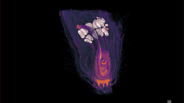
“This paper focuses on the multiscale,” says corresponding author Chris Topp, “because plants are multiscale. An ear of corn starts off as a microscopic group of cells called a meristem. Meristem cells will eventually form all the visible parts of the corn plant through division and growth.” Their improved 3D X-ray microscopy (XRM) technology allows the researchers to relate the developmental microstructure of the plant, such as meristem cells, to visible traits as they mature, for example leaves and flowers. In other words, 3D XRM provides cellular-level resolution of entire plant organs and tissues.
Read more at: Donald Danforth Plant Science Center
X-ray microscope 3D volume rendering of a developing soybean flower (Photo Credit: Donald Danforth Plant Science Center)